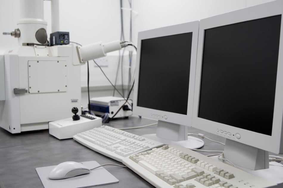
Image Credit: WH CHOW/Shutterstock.com
Applications of Automated Particle Analysis
Numerous problems can be solved by using automated particle analysis. The method can uncover what kinds of particles exist in a sample, the chemical composition of the detected particles, the particle size distribution of a powder mixture, and what rare materials are present in a mixture. It can also characterize the particle population without bias and correlate particle size and shape with composition.
The applications of automated particle analysis are numerous, diverse, and span a number of industries. The most common and traditional applications include its use in criminal forensics in analyzing gunshot residue, as well as conducting inclusion analysis to determine the cleanliness of steel and other metals.
There are also newer, and less common applications of automated particle analysis, such as the analysis of particles or contaminants in the hard disk industry, analyzing particles or contaminants in other devices with high-speed moving parts, and cleanliness and contamination measurements.
It can also be used for wear debris analysis, inclusions or constituents in aluminum alloys, analysis of inclusions in tissue, analysis of particles in pharmaceutical products, measure of wear metals from lubricating fluids, mineral grains and inclusions in other minerals, assessment of environmental particles from air or water samples in environmental studies, and the analysis of nano-particles.
While particle analysis has been established for a number of years, scientists have improved on the method. The use of scanning electron microscopy (SEM) and energy dispersive x-ray spectrometry (EDS) has established a highly accurate technique for automated particle analysis.
Using SEM and EDS to Automate Particle Analysis
The SEM technique uses an electron microscope that generates images of the surface of a sample by projecting a focused beam of electrons onto it. This interacts with the sample’s atoms, generating signals dependent on the sample’s characteristics which can be read and analyzed. EDS is used alongside SEM and is an advanced chemical microanalysis technique.
The combination of these two methods to conduct automated particle analysis has allowed multiple industries to answer questions surrounding particle populations in various scenarios.
The method takes advantage of the high-resolution imaging of SEM and the microanalysis capabilities of EDS along with the benefits of automation. This allows researchers to reliably analyze large populations of particles, from sizes of around 1,000 to 1,000,000 particles.
The process of SEM/EDS automated particle analysis first involves generating a high contact image to highlight the particles in the sample. An image analysis algorithm is then applied to differentiate between the distinct particles, allowing the analysis system to recognize the particles.
Next, the system records the location, size, and shape parameters of each particle in the sample. Following this, the microscope’s automated system reads the EDS spectrum of each particle, which generates a chemical analysis for different elements, such as iron and oxygen, to create different EDS spectrums.
The microscope moves over the material, conducting this analysis over different segments of the sample until a predetermined number of particles or fields of view have been analyzed.
At the end of the analysis, the results are tabulated to offer a complete profile of the particles present in the sample. Using this method, thousands of particles can be automatically characterized for a number of features in just a few hours. There is no requirement for a human worker apart from in the setup phase.
The Benefits of the SEM/EDS Method
SEM and EDS can be used together successfully to perform automated particle analysis. The combination of high-resolution imaging with microanalysis generates powerful datasets that describe numerous characteristics of the sample material.
The benefits of this method are that workers are not required to conduct the analysis, only set it up. Thousands of particles can also be studied simultaneously to generate data that helps professionals in various industries to solve numerous questions about specific materials.
References and Further Reading
Disclaimer: The views expressed here are those of the author expressed in their private capacity and do not necessarily represent the views of AZoM.com Limited T/A AZoNetwork the owner and operator of this website. This disclaimer forms part of the Terms and conditions of use of this website.