Feb 9 2016
It can be difficult to see everything necessary, and in great detail, during brain tumor surgery. Often, a neurosurgeon is choosing between breadth (microscope glasses known as “loupes”) and depth (a microscope sitting outside the surgical field) as the surgery goes on.
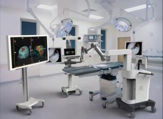
A new technology coming to our new operating rooms in July combines the two views into one, mounted on a robot. The whole surgical team will be able to see everything the surgeon sees, in high definition on a large monitor.
“This type of technology can change our perspective to further refine and improve how we do surgery,” says Karin Muraszko, M.D., chair of neurosurgery at U-M.
The technology, called BrightMatter, is from a company called Synaptive Medical we’ve been working with on research to improve brain surgery. We’ll use it to operate on brain tumors, aneurysms, vascular lesions and skull-based issues. U-M treats around 200 primary brain tumors alone each year, and we’re one of the first hospitals in the country to incorporate BrightMatter into our operating rooms.
“We’re creating the OR of the future, and defining the tools that will be a part of that,” says U-M neurosurgeon Daniel Orringer, M.D.
The long, automated arm of the device contains a scope and high-powered lighting that provides incredibly detailed vision to surgeons, from gross anatomy to fibers and cells. This arm is linked to sensors built into the surgeon's tools to inform the surgeon as to their location within the brain at all times.
Using images created during detailed pre-planning of the surgery, sensors let the doctors know if they are near critical anatomy within the patient's brain.
“Better imaging makes brain surgery safer and faster,” says U-M neurosurgeon Oren Sagher, M.D. “We’ll have more flexibility in July, once we can incorporate the image-guided technology we already have with our new technology.”
Always improving
Orringer’s stimulated Raman scattering (SRS) microscope is another way we’re continuing to improve imaging during neurological surgery.
To get microscopic images today similar to what the SRS microscope produces, surgeons have to wait a half hour or more for tissue to be frozen, sectioned, stained and interpreted by expert pathologists trained to spot the difference between cancer cells and normal brain cells.
“Our longer-term plans would be to use the BrightMatter device together with the SRS microscope,” Sagher says. “We hope to eventually replace microscopes with this type of device.”
Source: http://www.uofmhealth.org/