Image Credit: University of Cambridge.
All over the world, head injury is a huge public health burden and has an impact on around 60 million people each year. It is the major cause of mortality in young adults. A head injury patient is often sent for a CT scan to check for the presence of blood in or around the brain, and to help identify whether surgery is needed.
CT is an incredibly important diagnostic tool, but it’s rarely used quantitatively. Often, much of the rich information available in a CT scan is missed, and as researchers, we know that the type, volume and location of a lesion on the brain are important to patient outcomes.
David Menon, Study Co-Senior Author and Professor, Department of Medicine, University of Cambridge
Various types of blood in or around the brain can result in different patient outcomes, and usually, radiologists make estimates to identify the best course of treatment.
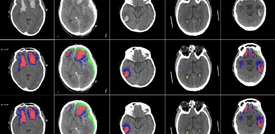
Detailed assessment of a CT scan with annotations can take hours, especially in patients with more severe injuries. We wanted to design and develop a tool that could automatically identify and quantify the different types of brain lesions so that we could use it in research and explore its possible use in a hospital setting.
Dr Virginia Newcombe, Study Co-First Author, Department of Medicine, University of Cambridge
The research team created a machine learning tool based on an artificial neural network. The tool was trained on over 600 different CT scans, illustrating brain lesions of various sizes and types. Then, the tool was validated on an existing huge dataset of CT scans.
The AI could perform classification of individual parts of each image and denote whether or not it was normal. In future studies, this could be useful to analyze how head injuries progress, as the AI may be more consistent compared to a human at finding minute changes over time.
“This tool will allow us to answer research questions we couldn’t answer before,” stated Newcombe. “We want to use it on large datasets to understand how much imaging can tell us about the prognosis of patients.”
We hope it will help us identify which lesions get larger and progress, and understand why they progress so that we can develop more personalised treatment for patients in future.
David Menon, Study Co-Senior Author and Professor, Department of Medicine, University of Cambridge
At present, the researchers intend to use the AI algorithm only for research purposes. However, they added that with proper validation, it could even be used in specific clinical scenarios, for example, in resource-limited areas with only a few radiologists.
Moreover, according to the researchers, it could be potentially used in emergency rooms, which will help patients return home sooner. Only 10% to 15% of all the patients who suffer a head injury have a lesion that can be viewed on a CT scan.
The AI could prove useful in identifying such patients who require further treatment, enabling those without a brain lesion to be sent home. However, any clinical application of the tool would have to be validated carefully.
The capability to automatically examine huge datasets will also allow the researchers to find solutions to crucial clinical research questions that have earlier been challenging to solve, such as the identification of relevant features for prognosis, which in turn may help target therapies.
The study was partially supported by the European Union, the European Research Council, the Engineering and Physical Sciences Research Council, Academy of Medical Sciences/The Health Foundation, and the National Institute for Health Research.
Journal Reference:
Monteiro, M., et al. (2020) Multiclass semantic segmentation and quantification of traumatic brain injury lesions on head CT using deep learning: an algorithm development and multicentre validation study. The Lancet Digital Health. doi.org/10.1016/S2589-7500(20)30085-6.