Digital pathology is a new field that mostly deals with microscope pictures obtained from patient biopsies. Due to their great quality, most whole slide images (WSI) are rather huge, often exceeding a gigabit (Gb). As a result, traditional picture analysis algorithms are unable to effectively manage them.
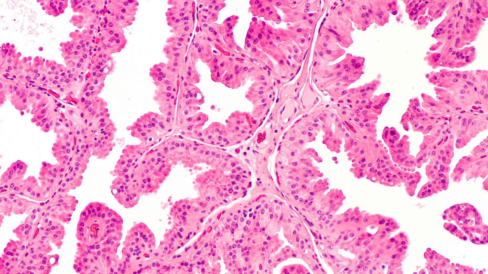
Image Credit: Shutterstock.com/ David A Litman
Researchers at Boston University School of Medicine (BUSM) saw a need for and built a revolutionary artificial intelligence (AI) technique based on a framework called representational learning to classify lung cancer subtypes from resected tumor pictures.
We are developing novel AI-based methods that can bring efficiency to assessing digital pathology data. Pathology practice is in the midst of a digital revolution. Computer-based methods are being developed to assist the expert pathologist. Also, in places where there is no expert, such methods and technologies can directly assist diagnosis.
Vijaya B. Kolachalama, PhD, Study Corresponding Author and Assistant Professor, Medicine and Computer Science, Boston University School of Medicine
The researchers created Graph Transformer (GTP), a graph-based vision transformer for digital pathology that uses a graph representation of pathology pictures and the computing efficiency of transformer designs to analyze the entire slide image.
Translating the latest advances in computer science to digital pathology is not straightforward and there is a need to build AI methods that can exclusively tackle the problems in digital pathology.
Jennifer Beane, PhD, Study Co-Corresponding Author and Associate Professor, Medicine, Boston University School of Medicine
Scientists then created a methodology that could differentiate between lung adenocarcinoma, lung squamous cell carcinoma, and adjoining non-cancerous tissue utilizing slide images as well as clinical data from three publicly available national cohorts. Researchers demonstrated that their GTP framework outperforms existing state-of-the-art approaches for full slide image classification in a series of trials and sensitivity evaluations.
Their machine learning methodology, researchers think, has implications beyond digital pathology. “Researchers who are interested in the development of computer vision approaches for other real-world applications can also find our approach to be useful,” researchers concluded.
IEEE Transactions on Medical Imaging published these findings online.
Journal Reference:
Zheng, Y., et al. (2022) A graph-transformer for whole slide image classification. IEEE Transactions on Medical Imaging. doi.org/10.1109/TMI.2022.3176598.