Image Credit: Chalmers University
New computer-aided techniques for reading medical images are being developed for a range of medical conditions. They can help radiologists by providing a second opinion or prioritizing patients who require care the quickest.
In close partnership with Sahlgrenska Academy at the University of Gothenburg and Sahlgrenska University Hospital, she has been actively involved in advancing medical imaging technologies within the realm of cancer, alongside numerous other medical conditions, including cardiovascular disease, stroke, and osteoporosis.
Large Study to Track Cancer in the Lymphatic System
Ida Häggström has recently worked with clinically active researchers at several institutions, including Memorial Sloan Kettering Cancer Center in New York, to develop a computer model published in The Lancet Digital Health.
Based on more than 17,000 images from more than 5,000 lymphoma patients, we have created a learning system in which computers have been trained to find visual signs of cancer in the lymphatic system.
Ida Häggström, Associate Professor, Department of Electrical Engineering, Chalmers University of Technology
Researchers looked through picture archives dating back more than a decade for this study. They contrasted the patients' final diagnosis with pre-and post-treatment computed tomography (CT) and positron emission tomography (PET) scans. The AI computer model was then trained using this data to identify indicators of lymph node cancer in an image.
Supervised Training
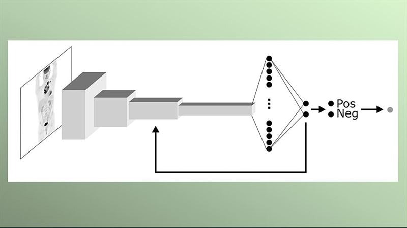
Ida Häggström created the Lymphoma Artificial Reader System, or Lars, a computer model that is referred to as a “deep learning system” based on artificial intelligence. To operate, an image from PET must be entered, and the AI model must then analyze this image. It is trained to look for patterns and characteristics in the picture to provide the most accurate estimate of the image’s positivity or negativity or whether or not lymphoma is present.
Häggström added, “I have used what is known as supervised training, where images are shown to the computer model, which then assesses whether the patient has lymphoma or not. The model also gets to see the true diagnosis, so if the assessment is wrong, the computer model is adjusted so that it gradually gets better and better at determining the diagnosis.”
What implications arise from the computer model employing artificial intelligence and deep learning for diagnosis?
Häggström stated, “It’s about the fact that we haven’t programmed predetermined instructions in the model about what information in the image it should look at, but let it teach itself which image patterns are important in order to get the best predictions possible.”
Support for Radiologists
Ida Häggström says the process of instructing the computer to identify cancer in images is time-intensive, noting that the study has spanned several years. Generating a significant volume of image data has posed a significant challenge. Adapting the computer model to differentiate between cancer and transient treatment-related alterations observed in images following radiotherapy and chemotherapy has also proven difficult.
Häggström added, “In the study, we estimated the accuracy of the computer model to be about 90 %, and especially in the case of images that are difficult to interpret, it could support radiologists in their assessments.”
However, a great deal of work must be done to validate the computer model if it is to be used in clinical practice.
Häggström concluded, “We have made the computer code available now so that other researchers can continue to work on the basis of our computer model, but the clinical tests that need to be done are extensive.”
Journal Reference:
Häggström, I., et al. (2024) Deep learning for [18F]fluorodeoxyglucose-PET-CT classification in patients with lymphoma: a dual-center retrospective analysis. The Lancet Digital Health. doi.org/10.1016/S2589-7500(23)00203-0