Image Credit: College of Engineering, Carnegie Mellon University.
However, this process is time-intensive and must be carried out by a specialist. Therefore, a majority of the placentas are not studied after birth.
A group of scientists from Carnegie Mellon University (CMU) and the University of Pittsburgh Medical Center (UPMC) has described a new machine learning method to study the placenta slides in the American Journal of Pathology, from Elsevier. The new method would enable more women to be aware of their health risks.
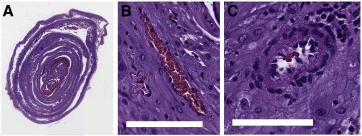
One of the reasons for studying the placentas is to look for a kind of blood vessel lesions known as decidual vasculopathy (DV). These lesions show that the mother is at risk of preeclampsia—a complication that could prove deadly to the mother and the baby in any later pregnancies.
Preeclampsia can be treated if detected in time, so there is a significant advantage from identifying mothers who are at risk before symptoms show up. But despite the fact that there are hundreds of blood vessels present in an individual slide, only one diseased vessel is required to pinpoint the risk.
Pathologists train for years to be able to find disease in these images, but there are so many pregnancies going through the hospital system that they don't have time to inspect every placenta. Our algorithm helps pathologists know which images they should focus on by scanning an image, locating blood vessels, and finding patterns of the blood vessels that identify DV.
Daniel Clymer, PhD, Alumnus, Department of Mechanical Engineering, Carnegie Mellon University
Machine learning “trains” the computer to identify some features in data files. In such a case, the data file turns out to be an image of a thin slice of a sample of the placenta. The team shows the computer several images and pinpoint if the placenta is healthy or diseased. Following sufficient training, the computer becomes capable of independently determining the diseased lesions.
It is very hard for a computer to just look at a huge image and sort it. Therefore, the researchers developed a novel method that can be used by the computer to track an array of steps to render the task more tangible.
Initially, the computer identifies all blood vessels that exist in an image. Then, every blood vessel can be examined separately, thus forming tiny data packets for the analysis. Next, the computer accesses each blood vessel and decides if it should be classified as healthy or diseased.
Moreover, at this point, the algorithm takes into account pregnancy features like birth weight, gestational age, and any other conditions that the mother might have.
If any diseased blood vessels exist, then the picture—and thus the placenta—is labeled as diseased. The UPMC researchers offered the de-identified placenta images to train the algorithm.
This algorithm isn’t going to replace a pathologist anytime soon. The goal here is that this type of algorithm might be able to help speed up the process by flagging regions of the image where the pathologist should take a closer look.
Daniel Clymer, PhD, Alumnus, Department of Mechanical Engineering, Carnegie Mellon University
“This is a beautiful collaboration between engineering and medicine as each brings expertise to the table that, when combined, creates novel findings that can help so many individuals,” noted lead investigators Jonathan Cagan, PhD, and Philip LeDuc, PhD, professors of mechanical engineering at CMU, Pittsburgh, PA, USA.
As healthcare increasingly embraces the role of artificial intelligence, it is important that doctors partner early on with computer scientists and engineers so that we can design and develop the right tools for the job to positively impact patient outcomes. This partnership between CMU and UPMC is a perfect example of what can be accomplished when this happens.
Liron Pantanowitz, MBBCh, Study Co-Author, Formerly Vice Chair for Pathology Informatics, University of Pittsburgh Medical Center
Journal Reference:
Clymer, D., et al. (2020) Decidual Vasculopathy Identification in Whole Slide Images Using Multiresolution Hierarchical Convolutional Neural Networks. American Journal of Pathology. doi.org/10.1016/j.ajpath.2020.06.014.